
18 FDG PET-CT:
Positron emission tomography (PET) is a cutting edge technology in the armamentarium of the diagnostic field of clinical practice, which demonstrates the biological changes occurring at the cellular level before anatomical changes take place.
PET-CT is a combination of a PET scanner and a High Resolution Multi-slice CT which offers a unique hybrid form of fusion imaging by combining the high resolution structural images of CT with the functional images of PET, which provides a more complete picture of disease status.
PET-CT imaging has revolutionized many fields of medical diagnosis including Oncology, Neurology, Cardiology, Surgical planning and Radiotherapy planning particularly in cancer management.
 This modality uses administration of 'positron emitting isotopes' which get trapped in specific target areas to accurately stage the disease process. One such radiopharmaceutical 18F FDG is a glucose molecule tagged to 18F radioisotope. Cancer cells utilize more glucose compared to the normal tissue and so by administering this radiotracer we can study the metabolic behaviour of cancer cells. These changes often predate the conventional radiological modalities and thus can diagnose cancers in early stages and also can predict the aggressiveness of malignancies.
In the field of Oncology PET-CT finds its use in every stage of management of patients, from diagnosis to staging to assessing treatment response and to differentiate between scarring and recurrent disease. It is now being increasingly used for defining tumor volumes for precise targeting and for guided metabolic biopsy.
PET-CT often serves a purpose of "one stop shop" for the cancer work up and on many occasions avoids the need of multiple investigations and imaging.
Other than Oncology, PET is highly useful in Cardiology to assess myocardial viability before the revascularization procedures like CABG or PTCA.
PET-CT is also useful in Neurology to detect seizure focus, diagnose dementias like Alzheimer's disease and Movement Disorders like Parkinson's disease and various Psychiatric disorders etc.
PET-CT can be used in infection/ inflammatory disorders like Tuberculosis, Sarcoidosis and Pyrexia of Unknown Origin (PUO).
FAQ
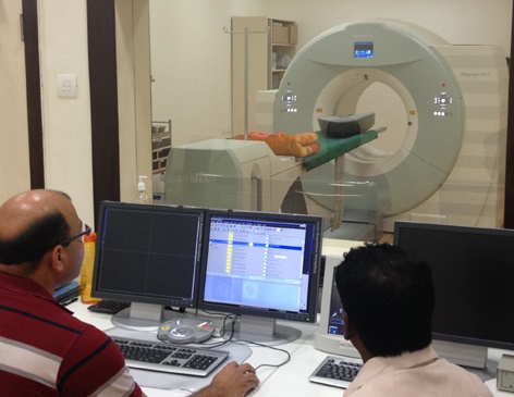
HOW DOES THE EQUIPMENT LOOK LIKE?
 A PET-CT scanner is a large machine with a round, doughnut shaped hole in the middle, similar to a CT or MRI unit. Within this machine are multiple rings of PET and CT detectors which will scan your body. You will lie on a narrow examination that slides into and out of this tunnel.
HOW DOES PET-CT WORK?
PET-CT is an advanced molecular imaging procedure that provides a way to diagnose disease through the measurement of metabolic activity. CT stands for Computerized Tomography. CT is an X-ray test that generates a detailed view of the structure of organs and tissues in the body. The use of oral and IV contrast agents can enhance the details by highlighting the gastrointestinal tract (filled by oral contrast) and other organs and blood vessels (filled with IV contrast).
During a PET scan, the patient is first injected with a radiopharmaceutical compound, usually FDG, a radioactive glucose compound. The compound distributes throughout the body and accumulates in various organs depending on the metabolic activity within the organ or tissue. Because cancer cells usually have a higher metabolic rate than surrounding cells, they utilize more FDG and will show up more prominently on the image. The PET scanner detects the FDG accumulated in glucose-avid organs or tissues and creates three-dimensional images of tissue function or cell activity in the tissues of your body that are displayed as colour-coded images. Even when an abnormal growth is not yet visible on a CT scan, the PET scan can show the abnormal cell activity as disease processes often begin with functional changes at the cellular level.
By combining the CT images and PET images, PET-CT gives complete structural and functional details of the disease in the whole body. By seeing these images, doctors can pinpoint the exact location of abnormal cell activity. They can also see the level and extent of that activity.
IS PET-CT SAFE PROCEDURE?
PET-CT is a safe procedure and millions of patients undergo worldwide. PET-CT is a non invasive and painless procedure with the exception of intravenous injection and is rarely associated with significant discomfort or side effects.
WHAR ARE THE BENEFITS AND RISKS OF PET-CT?
Benefits:
Nuclear medicine examinations offer information that is unique-including details on both function and structure and often unattainable using other imaging procedures. For many diseases, nuclear medicine scans yield the most useful information needed to make a diagnosis or to determine appropriate treatment, if any. By identifying changes in the body at the cellular level, PET imaging may detect the early onset of disease before it is evident on other imaging tests such as CT or MRI.
The benefits of a combined PET-CT scanner include:
Greater detail with a higher level of accuracy; because both scans are performed at one time without the patient having to change positions, there is less room for error. Greater convenience for the patient who undergoes two exams (CT & PET) at one sitting, rather than at two different times.
Risks:
PET-CT scans are very safe. You have a radioactive injection (tracer) and the CT scan uses radiation. But the doses of radiotracer administered are small, diagnostic nuclear medicine procedures result in relatively low radiation exposure to the patient, acceptable for diagnostic exams. The benefit of the scan, in finding out about your cancer, outweighs any potential risk from the radiation. Nuclear medicine diagnostic procedures have been used for more than five decades, and there are no known long-term adverse effects from such low-dose exposure. You will be informed of all significant risks prior to the exam and have an opportunity to ask questions.
Allergic reactions to radiopharmaceuticals may occur but are extremely rare and are usually mild. Nevertheless, you should inform the nuclear medicine personnel of any allergies you may have or other problems that may have occurred during a previous nuclear medicine exam. Injection of the radiotracer may cause slight pain and redness which should rapidly resolve.
Women should always inform their physician or technologist if there is any possibility that they are pregnant or if they are breastfeeding. You should not bring babies, young children or anyone who is pregnant to the scanning department. If you are breast feeding, you have to express enough milk beforehand to get your baby through the first 6 hours after the scan. This isn't because there will be radiation in the milk. It is because the mother shouldn't be holding the baby closely during the time the radiation is in her body.
If you have had sedative drugs as part of your scan you may be drowsy afterwards and it is best if someone can accompany with you.
HOW SHOULD I PREPARE FOR THE PET-CT SCAN?
Avoid strenuous exercise for a couple of days before your appointment. Have a low carbohydrate meal the night before your appointment. Do not eat or drink anything for at least 6 hours before the exam, except for plain water. If your doctor has told you to take your regular medicine, take it with plenty of water. You have to bring reports, films, and CDs of your previous procedures including X-Ray, USG, CT, MRI, pathology and any prior PET-CT scans.
Wear comfortable clothes to the test. Metal objects including jewelry, dentures and hairpins may affect the PET- CT images and should be left at home or removed prior to your exam. You may also be asked to remove hearing aids and removable dental work.
You have to bring Blood Urea and Creatinine report done a couple of days prior to your appointment.
Plan to be at the PET-CT imaging facility for 3 to 4 hours on the day of your appointment.
If you have diabetes, your blood sugar level must be below 150 mg/dl when you arrive at the testing.
If you are pregnant or think you might be, or if you are breast-feeding, tell your doctor or technologist before the exam.
WHAT HAPPENS BEFORE THE EXAM?
When you arrive at our facility, you will be asked to register at the reception. A doctor or someone from medical staff will review complete medical history and you may be asked to deposit reports, films and CDs of your previous investigations which will be returned to you after the exam. You will then go to the lab where a small finger prick will be done to test your glucose level. This test must be done prior to radioisotope injection as this information is necessary for the PET/CT exam. You may be asked to change into hospital gown if necessary.
Next, a small intravenous line (IV) will be placed in your arm and a safe amount of radionuclide will be given to you. When the radioactive material is injected into your arm, you may feel a cold sensation moving up your arm, but there are generally no other side effects Following the injection, you will rest on a lounge chair in a quiet room for 45 to 60 minutes while the radionuclide is distributed through your body. You will not feel any effects from this process. The doctor may request an IV contrast for the CT portion of the exam, so the IV will be left in until your exam is finished. You may be asked to drink oral contrast during this waiting period.
WHAT HAPPENS DURING THE EXAM?
You will be asked to empty your bladder, and then you will go to the scanning room. The PET/CT scanner is a machine that does PET and CT scanning in the same procedure. The technologist will help you onto the table. A scanning table will move slowly through the large opening of the scanner. You must be able to lie very still on the table for the entire test. Next you may receive the contrast enhancing agent through your IV. A brief sensation may move up your arm. You also may get a warm, flushed feeling; a taste of salt or metal in your mouth; or nausea for a few minutes. This is normal, but you should tell the technologist about these or other reactions. The technologist will go into a room behind a glass window. The technologist can see you at all times during scanning and may give you added instructions. You'll be able to talk to the technologist through an intercom during the scan. You should lie as quietly as possible. The scan is painless; you should not feel anything. Your scan will be completed in 20-30 minutes and a delayed scan may be taken if necessary.
WHAT HAPPENS AFTER THE EXAM?
After the exam, you will be asked to go back to the waiting room. After screening your scan by the doctor you will be abled to leave our facility. Though the FDG and contrast agent quickly leaves your body, you may be asked to drink plenty of water to expedite the process. You may resume your normal daily activity and diet.
HOW SOON WILL THE SCAN RESULTS BE AVAILABLE?
A Nuclear Medicine expert who has specialized training in PET-CT scans will interpret the images and write a report. You may collect the report after two working days.
QUESTIONS AND CONCERNS
Some anxiety before and during a test is normal. But a PET-CT exam should not be a fearful experience for you. Feel free to express any concerns about your PET-CT exam. Please ask the medical staff any questions you may have.
 Medical cyclotron
Medical cyclotron is a shelf shielded particle accelerator used to produce relatively short lived radioisotopes. In this cyclotron, different radioisotopes can be produced for e.g., 15O, 11C, 13N, 18F with half lives ranging from 2 mins to 109 mins. These isotopes after combining with various biomarkers for e.g., 18F FDG (FLUORODEOXY GLUCOSE) are used to detect early and accurate detection of various cancers, their recurrence and metastasis. Other than Oncology these isotopes are highly useful in Cardiology to assess myocardial viability and in Neurology to detect Seizure focus and diagnose Dementias and Movement Disorders.